July 10, 2023: Scientists have harnessed the power of deep-learning artificial intelligence (AI) to enhance the diagnostic capabilities of chest X-rays, particularly for assessing heart conditions. This groundbreaking approach offers a rapid and accurate means of evaluating heart function and detecting diseases.
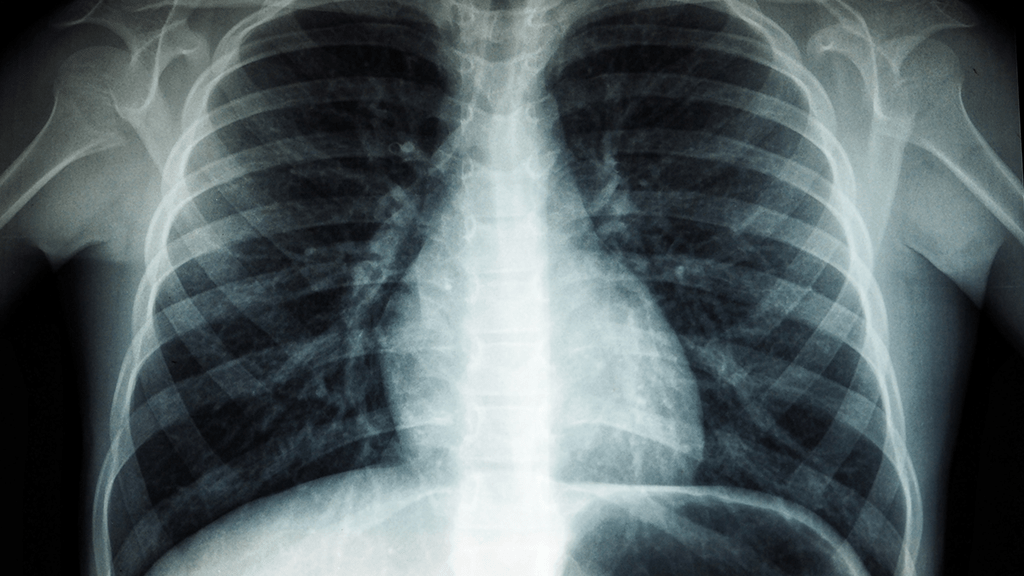
Healthcare professionals widely use chest X-rays to diagnose lung and heart ailments, making them the most commonly performed radiological test worldwide. While X-rays are convenient and quick, they provide only static images that lack information about how the heart is functioning. An echocardiogram, or “echo,” is needed to assess heart function.
An echocardiogram examines how effectively the heart pumps blood and identifies any issues with the heart valves, such as leaks or diseases. When the heart valves are diseased, the heart’s ability to pump blood efficiently is compromised, leading to heart failure, sudden cardiac arrest, or even death. However, performing an echocardiogram requires specialized skills and the presence of a trained technician.
Researchers from Osaka Metropolitan University have introduced a deep-learning AI model to transform chest X-rays into a more detailed diagnostic tool. Deep learning is an AI technique that enables computers to process data similarly to the human brain. The AI model generates accurate insights and predictions by recognizing intricate patterns in images, text, sounds, and other data.
The researchers trained the deep-learning model using a dataset of 22,551 chest X-rays and matching echocardiograms obtained from 16,946 patients across four facilities from 2013 to 2021. To ensure unbiased results, they incorporated data from multiple institutions.
The X-rays were input data, while the echocardiograms served as output data, allowing the model to learn the connecting features between both datasets. Testing the deep-learning model revealed its precise categorization of six types of valvular heart disease. The model achieved an area under the curve (AUC) ranging from 0.83 to 0.92 for differentiating between classes, with an AUC value closer to 1 indicating higher accuracy.
The researchers propose that their innovative AI approach could supplement echocardiograms, especially when quick diagnoses are required or when there is a shortage of technicians. This technology has the potential to benefit areas lacking specialist resources, respond to nighttime emergencies, and aid patients who face challenges in undergoing echocardiography.
Daiju Ueda, the study’s lead author, emphasized the significance of this research in improving the efficiency of doctors’ diagnoses and expanding access to diagnostic capabilities. While the study’s results required substantial time and effort, they offer promising advancements in the medical field.
Source: Osaka Metropolitan University